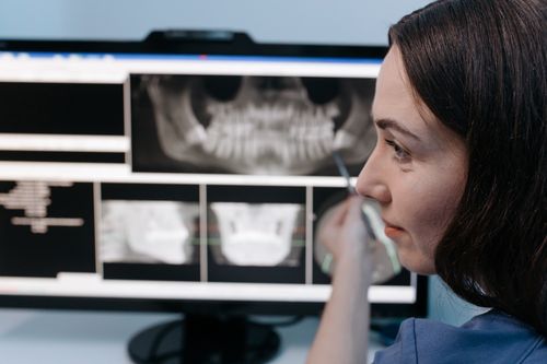
photo credit: Cottonbro Studio / Pexels
Key Takeaways
- Artificial intelligence enhances X-ray interpretation by helping radiologists detect abnormalities more quickly and consistently.
- AI can identify a wide range of pathologies, including lung diseases, fractures, cardiovascular conditions, and infections.
- Automated triage systems prioritize critical findings, helping clinicians respond faster to medical emergencies.
- AI serves as a clinical decision-support tool that improves workflow efficiency without replacing radiologists.
- As AI technology advances, it is expected to improve diagnostic accuracy, expand access to care, and support better patient outcomes.
Dr. Damon Deteso has worked as a private practice diagnostic radiologist with Millennium Medical Imaging in Saratoga Springs, New York, since 2004, where his responsibilities span emergency room imaging to routine outpatient studies across five area hospitals. He holds extensive experience with a range of imaging modalities, including computed tomography, magnetic resonance imaging, ultrasound, and X-ray, and he spent additional time as a medical advisor with Imagen Technologies training artificial intelligence systems focused on X-ray interpretation. Deteso earned his medical degree from the University of Massachusetts Medical School and completed his cross-sectional imaging training at the University of California, San Francisco. His academic foundation also includes a physics degree from Holy Cross University, where he was recognized by the Sigma Pi Sigma National Physics Honor Society.
That combined background in radiology and AI research offers a grounded perspective on how artificial intelligence tools are increasingly used to detect pathologies on X-ray images.
As one of the most commonly used diagnostic imaging tools worldwide, X-rays play a critical role in identifying a wide range of medical conditions. However, interpreting X-rays requires significant expertise and can be time-consuming, especially in busy health care settings.
AI-powered technologies are helping address these challenges by assisting radiologists in detecting pathologies quickly, accurately, and consistently.
One of the most common applications of AI in X-ray analysis is the detection of lung diseases. Chest X-rays are frequently used to evaluate conditions such as pneumonia, tuberculosis, chronic obstructive pulmonary disease, and lung cancer. AI algorithms can analyze subtle patterns within the lungs and identify abnormalities that may indicate disease.
AI has also demonstrated significant value in detecting lung nodules, which can be an early sign of lung cancer. Small nodules are sometimes difficult to identify, particularly when they overlap with normal anatomical structures. Advanced AI models can assist radiologists by flagging potential nodules for closer examination, potentially contributing to earlier diagnosis and improved treatment outcomes.
Fracture detection is another area where AI has shown considerable promise. Emergency departments frequently rely on X-rays to diagnose broken bones, but subtle fractures can occasionally be missed, especially during periods of high workload.
AI systems can analyze skeletal X-rays and identify fractures involving the wrist, ankle, hip, ribs, spine, and other bones. These tools can act as a safety net by alerting clinicians to possible fractures that require further review. Faster and more accurate fracture detection can improve patient care and reduce delays in treatment.
In musculoskeletal imaging, AI can also assist in identifying degenerative conditions such as osteoarthritis. By evaluating joint space narrowing, bone spurs, and other characteristic changes, AI algorithms can help assess disease severity and support treatment planning.
Quantitative measurements generated by AI may provide more objective assessments than traditional visual interpretation alone.
Cardiovascular abnormalities are another important target for AI-based X-ray analysis. Chest X-rays can reveal signs of heart enlargement, fluid accumulation in the lungs, and other indicators of heart disease.
AI systems can automatically assess cardiac size and detect findings associated with congestive heart failure. Early recognition of these abnormalities may help clinicians initiate treatment sooner and potentially prevent disease progression.
Conditions such as pleural effusion, pneumothorax, and pleural thickening can often be detected on chest X-rays. Pneumothorax, which occurs when air accumulates between the lung and chest wall, is considered a medical emergency in some cases.
AI-powered triage systems can automatically flag suspected pneumothorax cases and move them to the top of the radiologist’s worklist, helping ensure timely review and intervention.
AI models can recognize imaging patterns associated with bacterial infections and fungal diseases. In regions with limited access to radiologists, AI-assisted screening programs may help identify patients who require further diagnostic evaluation, improving access to care and supporting public health initiatives.
AI can also prioritize urgent findings through automated triage. When an X-ray contains evidence of a critical condition such as a collapsed lung or severe fracture, the AI system can immediately notify clinicians or move the case higher in the review queue.
This capability helps ensure that patients with potentially life-threatening conditions receive prompt attention.
As AI technology continues to evolve, its role in X-ray interpretation is expected to expand. By assisting in the detection of lung disease, fractures, cardiovascular conditions, infections, and other pathologies, AI is helping health care providers improve diagnostic accuracy, streamline workflows, and deliver better patient outcomes.
FAQs
How is artificial intelligence used in X-ray interpretation?
Artificial intelligence analyzes digital X-ray images to identify patterns that may indicate disease or injury. It can detect subtle abnormalities, highlight areas of concern, and assist radiologists by providing an additional layer of review.
Rather than replacing physicians, AI functions as a clinical support tool that helps improve efficiency and diagnostic confidence.
What types of conditions can AI detect on X-rays?
AI systems can help identify numerous conditions, including pneumonia, lung cancer, tuberculosis, chronic obstructive pulmonary disease (COPD), fractures, osteoarthritis, heart enlargement, pleural effusion, pneumothorax, and certain infections.
Many AI models are trained to recognize imaging patterns associated with these conditions, allowing clinicians to prioritize cases that may require immediate attention.
Can AI improve emergency care?
Yes. AI-powered triage systems can automatically flag critical findings such as collapsed lungs, severe fractures, or other urgent abnormalities and move those cases higher in a radiologist’s review queue.
This prioritization can shorten the time to diagnosis and treatment, particularly in busy emergency departments where rapid decision-making is essential.
Does AI replace radiologists?
No. AI is designed to complement – not replace – the expertise of trained radiologists. Final diagnoses and clinical decisions remain the responsibility of qualified medical professionals who interpret imaging findings within the broader clinical context.
By automating repetitive detection tasks and highlighting potential abnormalities, AI allows radiologists to focus more attention on complex cases and patient care.
What are the future benefits of AI in medical imaging?
As AI technology continues to evolve, it is expected to improve diagnostic accuracy, streamline radiology workflows, and expand access to imaging expertise, particularly in underserved regions with limited specialist availability.
Continued advancements may also support earlier disease detection, more objective assessments, and better patient outcomes through faster and more consistent image interpretation.
About Damon Deteso
Dr. Damon Deteso is a diagnostic radiologist who has practiced with Millennium Medical Imaging in Saratoga Springs, New York, since 2004, supporting five area hospitals with services ranging from emergency imaging to outpatient studies. He previously served as a medical advisor with Imagen Technologies, focusing on AI-assisted X-ray interpretation. He earned his medical degree from the University of Massachusetts Medical School, completed imaging training at the University of California, San Francisco, and holds a physics degree from Holy Cross University.