Dr. Sanjiv M. Narayan, MD, PhD, is a leading cardiologist and professor at Stanford University with extensive expertise in treating complex heart rhythm disorders. He is known for pioneering work that merges bioengineering with traditional medicine to create next-generation rhythm disorder treatments.
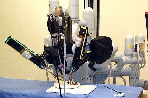
photo credit: Nimur / Wikimedia Commons. CC BY-SA 3.0
Dr. Narayan’s groundbreaking research in artificial intelligence (AI) and electrophysiology has influenced the development of innovative technologies, including AI-guided atrial fibrillation mapping systems now used by companies like Cortex Inc (formerly Ablacon). As co-Director of the Stanford Arrhythmia Center and Director of the NIH-funded T32 Program in Computational Medicine in the Heart, Dr. Narayan continues to lead advancements that improve cardiovascular outcomes and patient care.
Artificial Intelligence (AI) has changed various healthcare fields, with cardiovascular medicine benefiting as well. In this domain, AI helps tackle once-persistent challenges such as diagnosis accuracy and surgical safety and outcomes, as well as brings key improvements in different areas.
AI has streamlined cardiovascular disease and stroke imaging, largely circumventing past image processing and interpretation challenges. AI-powered tools make diagnosing and prognosis of cardiovascular conditions easier by automating abnormality detection, measuring plaque and blood flow, and predicting sudden cardiac death or stroke risk. AI also offers quick automated measurements for procedures, cutting down treatment time. Still, institutions using AI for medical imaging should be cognizant of the challenges tied to acquiring data and organizing and labeling it, as well as address ethics, bias, and data quality concerns.
Electrocardiography (ECG), a medical test tracking heart rhythm, has also changed with AI. With too few experts to interpret the growing number of ECGs, AI offers a solution by automating this task. Current ECG interpretation devices are rule-based and have limits. However, AI and machine learning (ML) can read ECGs more like human experts, leading to better care decisions. Sometimes, AI can read ECGs better than specialists, spotting hidden heart problems and finding conditions like aortic stenosis (narrowing of the heart’s aortic valve opening), something traditional methods often miss.
When paired with photoplethysmography (PPG), light-based method to track blood flow in the skin, AI can spot atrial fibrillation (AF), an irregular heartbeat. Smartwatches and other wearables with PPG can catch AF in real time, even outside clinical settings. By analyzing the timing and shape of the PPG signal, AI algorithms can differentiate between AF and a normal heart rhythm. This setup is more efficient than older methods like regression models and manual feature extraction, which lack precision. Complex AI models can also automatically find critical patterns in the PPG signal.
In hospitals, AI and machine learning boost bedside monitoring. For years, bedside monitoring has relied on pre-set rules to trigger alarms when a patient’s vital signs exceed a specific range. But these systems often give false alarms and look at each vital sign in isolation, often missing the connections between them. AI-based tools can simultaneously watch all the data from monitors and see subtle patterns across many vital signs. This approach gives a fuller picture of patient health and reduces false alarm detection.
Research highlights ways AI improves hospital monitoring beyond sorting true from false alerts. For instance, AI models can spot small physiological changes indicating a patient is getting sepsis, where the body overreacts to infection. AI tools also help doctors act quickly by predicting when a patient might experience cardiac arrest in the hospital. Moreover, they assess surgery risks by weighing various patient factors before, during, and after procedures, predicting complications and mortality, and guiding anesthetic drug dosing.
AI helps researchers and doctors study heart disease genes to create better treatments. For example, genome-wide association studies (GWAS) use AI to scan the entire genome, spotting patterns and links between genes and specific heart conditions. AI and machine learning methods such as gradient boosting sharpen polygenic risk scores (PRS), which gauge a person’s genetic odds of developing cardiovascular disease. Training AI on diverse, multi-ethnic genomic data makes these models work better for different groups. AI also connects observable traits, or phenotypes, to genes by analyzing facial images to detect genetic syndromes tied to heart issues.
Teamed with intelligent robots, AI boosts precision in complex surgeries while keeping them less invasive. The da Vinci Surgical System, for instance, enables robot-assisted surgeries, reducing risk and shortening hospital stays. For heart procedures like coronary interventions, AI robots let doctors work from a safe distance, avoiding radiation exposure.